Describe briefly the structure and development of the sporophyte of Marchantia, and add a brief note that how the young gametophyte develops from the sporophyte.
Q. Describe briefly the structure and development of the sporophyte of Marchantia, and add a brief note that how the young gametophyte develops from the sporophyte.
Or, Describe the post fertilization stages in Marchantia.
Ans. Zygote or fertilized egg is the first cell or unit of sporophyte of Marchantia. After fertilization the archegonium enlarges, due to which the sporophyte is continually enclosed within the archegonium, which ultimately forms a protective sheath-the calyptra. In addition the surrounding gametophytic tissue and cells below the archegonium also get stimulus, divide and redivide and form a cylindrical out growth the pseudoperienth. Thus the mature marchantia sporophyte has three protective coverings-a calyptra, pseudoperienth and involucre.
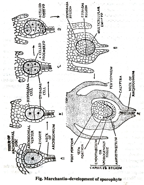
At the time of development the zygote increases in size (Fig.) and fills the cavity of venter, and secretes a wall around it. The zygote first of all divides by a transverse division into two cells (Fig.) the upper hypobasal and lower epibasal. Now both the cells further divide by a vertical wall forming quadrant stage (Fig.). The two upper hypobasal cells give rise to foot and seta, while the epibasal half develops into capsule. The four celled zygote or embryo further divides by a vertical wall at right angles to each other forming octant stage or 8 celled embryo. Now the embryo elongates and further divides by several irregular divisions so as to form a multicellular globular embryo. This multicellular embryo get differentiated into foot cell, seta cells (from hypobasal region) and capsuler cells from epibasal region. Now the sudden increase in length of the seta takes place by an elongation of its cells. Growth beyond such enveloping structure as involucre and pseudoperienth. Cell divisions in development of a foot are at various angles to one another. When mature, the foot may be bulbous as in M. polymorpha. The foot serves as an organ for anchorage and for absorption of food from adjoining gametophytic tissues. However the presence of some chloroplasts shows that sporophyte is not wholly dependent upon the gametophyte for the products of photosynthesis. The capsuler part of embryo soon divides by periclinal division so as to form a outer amphithecium and inner endothecium. The amphithecium forms the wall of capsule and endothecium forms archesporium which in turn forms the sporogenous tissue and spores. The archesporium divides and redivides and forms the sporogenous tissue. The sporogenous tissue get separated from elaters, while rest of sporogenous cells divide transversally to form vertical rows of spore mother cells or sporocytes. The spore mother cells divide by a reduction division (meiosis) and followed by a simple division to form four spores from each spore mother cell. The spores are haploid in nature.
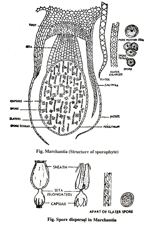
The mature sporangium is a elongated structure differentiated into definite foot, seta and capsule. The foot is a bulbous structure towards the base of archegonium, it helps in absorption and anchoring. The seta is short, thick stalk connecting foot and capsule. The capsule is a spherical structure at the distal end of sporogonium. It has a sterile jacket layer of cell. Inside the wall there are spores and elaters. The elaters are long, narrow, spindle-shaped cells pointed at each end, with two spiral thickenings on their walls. These elaters are hygroscopic in nature. The spores are spherical haploid structure with two layered wall-exine and intine. The exine being thick and warty. Inside the walls of spores there is a haploid nucleus and cytoplasm along with some chloroplasts.
Dispersal of spores and dehiscence of the capsule :
At maturity the wall of capsule get ruptured by irregular clefts due to the hygroscopic movements of elaters. After rupturance of wall of sporophyte the spore get liberated free and taken by air.
The germination of spore and development of gametophyte :
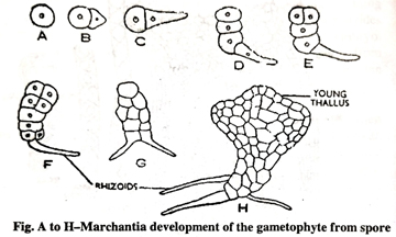
The spore is the first cell of the gametophyte. The spores are spherical and very small structures having a spore coat of endospore and exospore. The exospore or exine is thick and warty while endospore is thin. Inside the wall of spore a single nucleus with granular cytoplasm.
The spores just after dispersal on getting the suitable conditions starts germination. The spores increase to size and its exospore ruptures and endospore comes out in a form of short filaments. The filament like structure by division and redivisions forms the multicellular structure and finally it get changed into dichotomously branched young gametophytic thallus of marchantia.
Follow on Facebook page – Click Here
Google News join in – Click Here
Read More Asia News – Click Here
Read More Sports News – Click Here
Read More Crypto News – Click Here