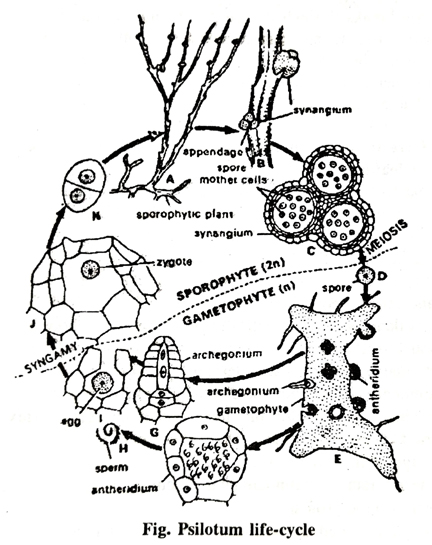
Describe the life cycle of Psilotum.
Q. Describe the life cycle of Psilotum.
Or, Describe briefly the life cycle of any homosporous pteridophyte studied by you.
Ans. The vegetative reproduction takes place by means of gemmae. These vegetative structures develop both on rhizomes (sporophyte) and prothalli (gametophyte).
(a) Gemmae on Sporophyte: The gemmae develop freely on the rhizomes. Each such gemma on being detached develops into a new rhizome. In starved conditions the upper cell of many of the rhizoids developed on the rhizome divides and produces a small nodular gemma. These gemmae are small oval bodies one cell in thickness. The cells remain filled with starch.
(b) Gemmae on Gametophyte: The gemmae also develop on the surface of the prothallus. In structure these gemmae resemble those which have been developed on the rhizome. These gemmae on being detached develop into prothalli.
The Synangium
Spore Producing Organs-The Synangia : The sporangia are borne in traids on minute appendages subtended by a bract. The sporangia remain fused with one another, and therefore, the group is called a synangium. The so-called synangium is generally interpreted as a sporangiophore fused with a subtending bract. The sporangia seem to be borne on the adaxial side of the appendage at the point of dichotomy and are slightly raised on broad short stalks. The slender branches of the fertile appendage are more or less erect and embrace the synangium. The fertile appendages are mostly grouped on the upper part of the stem, but both these regions are not strictly limited and sterile appendages may occur here and there among the fertile ones.
Structure of Mature Synangium: The synangium is three lobed when examined from the outside internally, it remains divided into three chambers. The wall is composed of several layers of cells. The sporangial wall cells have thickened considerably, except along one vertical line running from the distal end to the base of each sporangium. These are the lines of dehiscence, along which the synangium opens into three segments releasing spores. There is no true tapetum. The spores within the sporangium are bean-shaped with finely reticulated walls.
Development of Synangium: The development is apparently of the eusporangiate type. The earliest stages of development have shown, that each loculus arises separately from a single epidermal cell of the sporangiophore. It shows, that this structure is a union of three sporangia, the synangium. The primary initial divides periclinally separating a jacket initial and an archesporial initial. The jacket initial gives rise to a wall three to five cells in thickness, while the archesporial initial produces a central mass of sporogenous cells. Unlike most other pteridophytes, neither the outermost sporogenous cells nor the innermost jacket cells develop into a tapetum. The three sporangia are grouped around a central sterile tissue, the sporangiophore axis.
Division and redivision of the archesporial cell produce a large number of sporogenous cells. As the sporogenous tissue matures, some of the sporocytes are disorganized forming fluid that nourishes the surviving mother cells and spores develop. The spores are bean shaped with finely reticulated walls. Simultaneously the cells of the wall of sporangium become thickened, except along one vertical line from the apex to the base of the sporangium; these act as the lines of dehiscence.
The Gametophyte
Spore and its Germination: The spores are of equal size. Usually the spores are bead-shaped but some of them are found to be of the tetrahedral type. Each spore possesses a narrow ridge that joins the two ends of the curve. There is a median slit extending for about three fourth of its length. The surface spore is delicate and finely reticulate.
The spores germinate after about four months by rupture of its wall along the median slit and the spore contents project as a small protuberance covered with a thin membrane, the intine. Later on the extruded portion is cut off by a transverse wall so that the basal portion remains within the spore wall as a large spherical cell. The apical cell divides by oblique walls and a club-shaped filament is resulted. An apical meristem is established which apparently continues growth indefinitely resulting in a cellular body. This elongates into a cylindrical, slightly branched prothallus, covered with brown rhizoids. The prothallus grows by means of apical meristem. At this stage it closely resembles a portion of the rhizome. The prothallial tissue is colourless, saprophytic and mycorrhizal as in the sporophyte but it lacks vascular tissue, though cases are on record in which tracheids were found in large prothalli.
The gametophyte is monoecious that it bears both antheridia and archegonia. The sex organs are irregular distributed on the prothallus. The number of archegonia is lesser than that of antheridia.
Development of Antheridia: The antheridia appear first. They are prominently projecting, spherical bodies, with a wall of single layer of cells.
The antheridium arises from a superficial cell of the gametophyte. This cell also as the autheridial initial. The antheridial initial divides periclinally into an outer cell, the jacket initial, and an inner cell, the primary androgonial. The primary androgonial cell divides and redivides forming a considerable number of androgonial cells, the last generation of which are androcytes. The androcytes metamorphose into spirally coiled multiflagellate antherozoids. In the mean time jacket initial also divides anticlinally forming a jacket layer one cell in thickness which lies outside the outer face of androgonial tissue. There lies a triangular opercular cell in the centre of the antheridium which by its disintegration provides an opening for the escape of antherozoids.
Archegonium and its Development: The archegonium develops from a superficial cell of the gametophyte. This cell is known as archegonial initial. First of all this initial divides periclinally forming a primary cover cell and a central cell. Thereafter the primary cover cell divides anticlinally twice giving rise to four quadrately arranged neck initials. Now these neck initials divide periclinally forming an archegonial neck four to five cells in height and composed of four vertical rows of cells.
In the mean time the central cell also divides periclinally forming a primary canal cell and primary venter cell. As the neck develops the primary canal cell elongates vertically. According to Holloway and Lawson the division of neck canal cell is uncertain but it divides and gives rise to two neck canal nuclei which disintegrate later on. The behaviour of the primary venter cell is also doubtful. It is reported that it functions directly as the egg does not undergo the usual division forming venter canal cell and egg.
As the archegonia approach maturity, the cell walls of the lowermost tier of neck cells become cutinized and thickened. With the result the sloughing off the upper portion of the neck takes place. Simultaneously the neck canal cell or cells also disintegrate bearing a passage way for the entrance of antherozoids in the venter of the archegonium. One of the antherozoids penetrates the egg and the fusion of male and female nuclei takes place forming the oospore.
Development of Embryo : The development of embryo is very simple. The fertilized egg enlarges downward. It divides periclinally and the upper cell develops into the axis, the lower cell into a foot, which sends out fingerlike projections into the prothallial tissues. No root or cotyledon is formed. There is a marked line of division between axis and foot. When the developing axis breaks through the surface of the prothallus the foot and axis separate, the foot remains attached to the prothallus.

The first division in development of the epibasal cell into a foot is vertical. Thereafter the divisions take place in an irregular sequence in the two halves of the foot. As development of the foot continues it becomes a cylindrical structure of equal length and breadth. Superficial cells of the foot elongate into projections that enter the prothallus like haustoria.
The development of shoot takes place as follows: The epibasal daughter cell of the zygote divides vertically. The two daughter cells thus formed divide transversely. Subsequent divisions take place in irregular way. The short portion of a young embryo is hemispherical. It is differentiated vertically into two halves, derived, respectively from the first two cells of the shoot. The axis elongates vertically before it becomes free and develops an apical cell in one or in both halves of the young shoot. The apical cell always lie in the middle between base and apex of the half in which it is formed. Further development of the shoot takes place because of the activity of the apical cell or cells. When there are two apical cells the axis is dichotomous from the beginning, but in any case the first dichotomy soon takes place. In the early stages of the development of embryo the prothallus tissue also grows up like a calyptra round the young axis, but the growth activity of the embryo soon breaks through it. The protruding portion of the sporophyte soon develops rhizoids on its surface and becomes infected with a mycorrhizal fungus. Now this becomes a new independent plant. The rhizome continues to branch either dichotomously or by the formation of adventitious side branches. The original branch or some of the adventitious branches later on grow above the soil and develop into aerial shoots.
Follow on Facebook page – Click Here
Google News join in – Click Here
Read More Asia News – Click Here
Read More Sports News – Click Here
Read More Crypto News – Click Here