Describe the stem structure and reproduction of Rhynia.
Q. Describe the stem structure and reproduction of Rhynia.
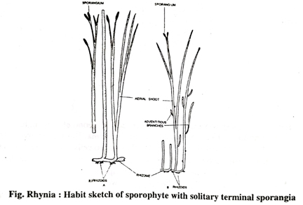
Ans. Aerial stem : The upright aerial shoot bearing no leaves arose from the rhizome, tapering from base upwards. The solid cylindrical greenish stem was both simple and dichotomously forked once or twice. The stem or branch tip was either sterile or terminated with a sporangium being slightly broader than the adjoining vegetative axis.
The adventitious branches meant for vegetative propagation were studded with the proximal end of the stem in R. Gwynnevaughani. They, being hemispherical and having own vascular strand, had no connection with the stem strand, and thus they are of exogenous origin (Fig.).
Stem anatomy: Both the rhizome and aerial stem were anatomically similar, except stomata and photosynthetic cortical cells which were absent in rhizomatous stem.
Epidermis was cuticularised uniseriate parenchymatous bearing stomata. Cortex: It was divisible into 2 distinct zones: (a) Outer cortex was single-layered thin-walled parenchymatous without any intercellular space; and (b) Inner cortex being multilayered comprising thin-walled spherical or oval cells that were loosely-placed owing to presence of sufficient air spaces.
The aeriferous system of this zone might be helpful in keeping the slender aerial stem vertically erect. A few peripheral layers of this zone might have been chlorophyllous.
Endodermis and Pericycle-Absent.
Vascular cylinder: It was haplostelic protostele, since the smooth core of solid xylem was completely surrounded by phloem; the haplostele is regarded to be the most primitive among protostele as this can serve the purpose of slender stem axis only. The protoxylem elements were exarch.
The xylem was mainly comprised of tracheids having annular thickenings on their radial wall. The phloem consisted of elongated elements with oblique end walls. It was 4-5 cells in radial extent.
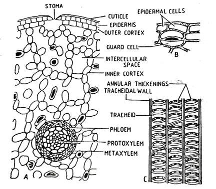
Fig. Rhynia: A. T.S. or a sector of stem; B. A stoma in surface: view; C. L.R.S. of xylem
Reproduction: The sporangia were cylindrical and borne singly on the tips of some aerial branches. They were large, oval or cylindrical structures with pointed ends. The sporangia had walls several cell layers thick in which the cells of the outermost layer were thick walled and had a heavy cuticle. The sporangium was without any specialised mechanism of dehiscence. The inner cells of the jacket probably functioned as tapetum. The sporangia contained numerous spores with cutinised walls. The spores were apparently all alike and were arranged in tetrads. The presence of tetrads in some specimens suggests that meiosis occurred in Rhynia and that the plant body, therefore, belonged to the sporophytic generation.
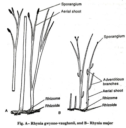
The Spores: The spores were cutinised and usually were borne in tetrads. Nothing is known about the gametophyte of Rhynia because the gametophyte of this fossil plant has never been discovered. Lyon (1957) reported some germinating spores from Rhynie Chert, which show multicellular structures, developing at the ends of germ tubes. These may represent the gametophytic generation. Merker (1959) has suggested that the underground parts of Rhynia might possible be the gametophytes. According to Pant (1961). R. gwynnevaughanii may be the gametophyte of the larger R. major.
Follow on Facebook page – Click Here
Google News join in – Click Here
Read More Asia News – Click Here
Read More Sports News – Click Here
Read More Crypto News – Click Here