Describe the structure and life history of Polysiphonia.
Q. Describe the structure and life history of Polysiphonia.
Ans. Systematic Position:
Class : Rhodophyceae
Sub-class : Florideae
Order : Ceramiales
Family : Rhodomelaceae
Sub-family : Polysiphonaceae
Genus : Polysiphonia
Occurrence: It is a common red alga on sea-coasts. It is found in Atlantic and Pacific Oceans. It is also found in India and Anand reported P. platycarpa, P. ferulacea and P. variegata from Karachi (Pakistan). The later species, according to Fritsch, is common in polluted water and roots of Mangroves. P. urceolata is an epiphyte on Laminaria, P. elongata is a lithophyte and is nearly 30 cm in length. P. fastigilata is a semi-parasite on Ascophyllum nodosum although Rattaray has reported the same species growing on stones in an independent state. But it is said that the species growing independently are not so healthy as those growing on the brown sea-weed.
Structure: The plants of Polysiphonia exhibit a variety of colours. They may be red, brownish red or even darker in colour. The plants form thick tufts and are attached to the substratum by means of long unseptate rhizoids. This thallus is laterally and dichotomously branched and is made up of filaments placed parallel to each other. They are called siphons and if seen in a microscope, the cells of the central siphon look large and elongated. On either side of the central siphon are situated pericentral siphons which are narrow. The number of siphons varies from four to twenty. These pericentral siphons in older branches may further cut off smaller shiphons on either side which are called cortical siphons. However, the tip of the thallus always consists of the cells of the central siphon. Some pericentral cells cut off small branches from their upper ends. They are of limited growth, monosiphonous and are called trichoblasts. The cells are connected by protoplasmic strands.
Each vegetative cell has a single nucleus, usual chromatophores and floridean starch as food material.
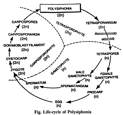
Reproduction: It is vegetative, sexual and asexual.
Sexual reproduction: The sexual reproduction is oogamous and is brought about with the help of male, female and tetrasporic plants. The male and the female organs of reproduction are known as antheridia and carpogonia respectively.
Development of antheridia: They develop on male plants in clusters near the tip of the main filament. Generally, a single cluster develops on a trichoblast but occasionally two clusters may develop as in P. platycarpa. In the beginning the filament developing antheridium is monosiphonous but very soon it becomes polysiphonous. Sooner or later as a result of these divisions a central axis of elongated cells is differentiated, surrounded by pericentral cells. From these pericentral cells, primary antheridial mother cells are developed which in their turn cut off secondary antheridial cells as in P. platycarpa (Smith has not reported the presence of such secondary cells in P. flexicaulis). So each pericentral cell bears a number of antheridial mother cells. According to Grubb and Kylin these mother cells bear two, three or four antheridial cells, depending upon the species. The formation of antheridial cells is very regular in P. platycarpa. Iyeongar and Balkrishnan have given a simple, clear and well illustrated account of their development. According to these workers, the second antheridium is cut just opposite to the first. Third and the fourth are cut in between first and the second.
So far as the development of antheridium is concerned, it is more or less identical in the different species of Polysiphonia. The whole male cluster is surrounded by a gelatinous investment. From the antheridia, spermatia are liberated, which are spherical or ovoid, uninucleate, non-motile with a delicate wall.
Development of carpogonium : It arises from a fertile pericentral cell. This cell divides into two-an upper carpogonial branch initial and a basal cell which is called bearing cell or supporting cell. The carpogonium branch initial further divides and thus a row of three cells is formed. So carpogonial branch in P. platycarpa is three celled whereas in P. violaceas and P. nigrescens it is four celled. The uppermost cell of the carpogonial branch now elongates and forms trichogyne while its basal region brodens into carpogonium where the female nucleus is located.
Pre-fertilization changes There are certain developments in the carpogonium before fertilization. The basal cell or the bearing cell cuts off a cell towards its base which is called the basal sterile cell. Soon afterwards another sterile cell is cut towards the side of the bearing cell which also divides into two. These two cells are called the sterile cells. The prefertilization developments in the Indian species P. platycarpa is identical upto this stage.
Fertilization: In some species like P. violacea the carpogonium nucleus divides at the time of fertilization and one of the nuclei passes into the trichogyne. However, the division of the carpogonium nucleus has not been observed in P. nigrescens.
At the time of fertilization, the spermatium adheres to the trichogyne. Finally, the wall between the two dissolves. More than one spermatium has also been noticed attached to the trichogyne in case of P. platycarpa. The nucleus of the male then passes downwards through the trichogyne into the carpogonium. The two nuclei unite resulting in the formation of a zygote.
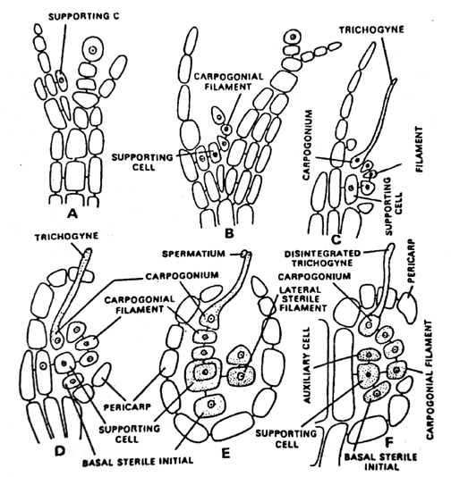
Fig. Polysiphonia, Sexual Reproduction. A. longitudinal section of the terminal part of the thallus before the development of carpogonial filament; B. developing carpogonial filament on the supporting cell; C. carpogonial filament; D. basal sterile initials cut off; E. lateral sterile filament cuts off; F. post-fertilization stages and development of auxiliary cell
Post-fertilization changes : After fertilization, many changes take place within and around the carpogonium. First, the basal sterile cell divides and thus two basal sterile cells are formed. The lateral sterile cells also divide and thus many lateral sterile cells are formed.
The bearing cell now enlarges and cuts off a cell above which is an important cell and is called the auxiliary cell. In P. platycarpa the auxiliary cell cuts off another small cell known as the connecting cell because it connects the carpogonium to the auxiliary cell. Such connection is, however, lacking P. nigrescens. The zygote nucleus now divides by mitosis and one of the nuclei passes into the auxiliary cell. The carpogonium is now cut off by a wall and finally degenerates. During all these changes, the bearing cell, the auxiliary cell and the sterile cells become interconnected and open communications are established between them. The function of the sterile cells is supposed to be nutritive. All these cells are so integrated to form a central mass called the fusion cell or the placental cell.
The fusion cell now cuts off the gonimoblast mother cell or gonimoblast initial. The zygote and nucleus also divide and one of the nuclei passes into the gonimoblast mother cell. The latter immediately cuts off a number of gonimoblast cells. Every time a new cell is cut, the zygote and nucleus divide and its derivative enters the newly formed cell. These gonimoblast cells at later stages are known as carpospores. All these carpospores are enveloped within the wall of the fruit body which is called cystocarp or carposporophyte. The wall of the carposporophyte is double layered in the beginning in P. platycarpa but as the development proceeds, the wall becomes single layered. The inner layer shrivels and finally disappears thus indicating its nutritive value. The carposporophyte has a pore.
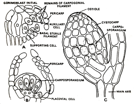
Fig. Polysiphonia, Post-fertilization stages. A. procarp after formation of first gonimoblast initial; B. Carposporophyte with carposporangia; C. surface view of mature pericarp (cystocarp)
The carpospores are finally liberated through this pore and on reaching a suitable place they germinate. According to Lewis they give rise to tetrasporic plants. As they are diploid in nature, the plants developed as a result of their germination are also sporophytes although apparently they resemble the normal gametophytic plants.
Follow on Facebook page – Click Here
Google News join in – Click Here
Read More Asia News – Click Here
Read More Sports News – Click Here
Read More Crypto News – Click Here