Give an illustrated account of the structure and life history of Vaucheria.
Q. Give an illustrated account of the structure and life history of Vaucheria.
Or, Describe the life history of Vaucheria and discuss its systematic position.
Or, Give a brief account of sexual reproduction in Vaucheria.
Ans. Systematic Position :
Class – Chlorophyceae
Order – Siphonales
Family – Vaucheriaceae
Genus – Vaucheria.
Occurrence : Vaucheria is a green filamentous freshwater alga found generally in spring water. Some species are terrestrial and occurred in damp and muddy soil in yellow colour. Some species like V. amphibia are found both on soil and in water. Some species are marine and found in sea water. The common species are V. germinata, V. sessilis, V. amphibia and V. uncinata.
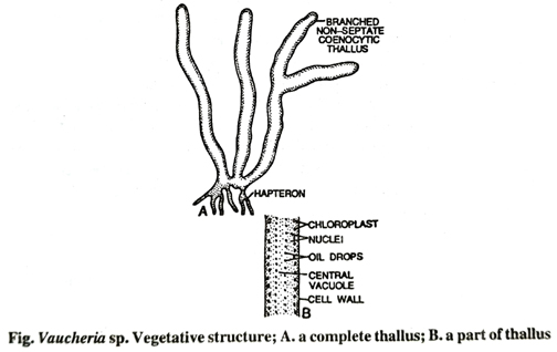
Structure: The plant body or the thallus is filamentous, profusely branched, coenocytic (multinucleate), unseptate and cylindrical. The filaments are tubular or siphonous and contains cytoplasm, chloroplast and several nuclei.

The thallus is attached to the substratum with the help of branched rhizoids or hapteron. The wall of the filament is made up of pectose. A central elongated vacuole is found throughout the plant body. The cytoplasm is found along the wall and contains many disc-shaped chloroplasts, several nuclei and oil droplets as reserved food. The pyrenoids and starch grains are completely absent.
Reproduction: The reproduction in vaucheria takes place by following methods:
1. Vegetative method: By fragmentation: The filament breaks into several pieces due to external disturbances. All pieces develop into new plants.
2. Asexual reproduction: The asexual reproduction takes place by means of (i) Zoospores (ii) Aplanospores (iii) Hypnospores.
(i) By zoospores: In aquatic species normally the zoospores are produced as coenocytic, compound, multiflagellate, spherical or oval in shape. They are formed singly in a zoosporangium. The zoospores are also called Synzoospores or Coenzoospores.
Formation of zoospores: Any branch of the filament becomes swollen at the apical end and functions as sporangium. The protoplast of the filament comes to the swollen end of filament, the vacuole disappears and the swollen end becomes club-shaped. A transverse septum appears below the swollen end which separates this from the rest filament. The swollen end is the sporangium. The rearrangement of protoplast takes place in which all the nuclei come towards the periphery. The protoplast contracts slightly and all the nuclei produce two flagella towards outside. Thus the whole protoplast of the sporangium metamorphosed into a multi-flagellate zoospore.
Liberation of zoospore: When the zoospore gets maturity a pore appears at the apical portion of the sporangium. The zoospore escapes through this pore into water.
Germination of zoospore: The zoospore after liberation comes into water and swims for some time and then comes to rest at the bottom. Now the zoospore withdraws the flagella and secretes a cellulose wall around itself. At this stage the rearrangement of protoplast again takes place, as a result the nuclei comes to the centre and the chloroplast goes towards the periphery. The zoospore begins germination giving rise to two germ tubes. One of them forms rhizoid and the other (upper) forms the plant body.
(ii) By aplanospore: The terrestrial species produce aplanospore in normal condition but the aquatic species produce it under unfavourable condition. The aplanospore is a non-flagellate, non-motile zoospore and formed in an aplanosporangium like zoospore. The protoplast comes to apical portion of any filament which becomes swollen. The whole protoplasm rounds off forming a spherical or oval aplanospore. A transverse wall separates the aplanosporangium from the rest of the filament. The aplanospore is thick walled and gets liberated through a pore appearing at the apex of the aplanosporangium.
The aplanospore germinates directly by producing 1-2 germ tubes which develop into a new plant thallus.
(iii) By hypnospore: Under unfavourable conditions the protoplast of the filament is divided into several pieces due to segmentation of septation. All segments are multinucleate which secrete a thick wall around themselves. The coenocytic thick walled bodies are called hypnospores. These hypnospores are arranged linearly in such a way that they resemble like Gongrosira algae. Therefore this stage is called the Gongrosira stage. The hypnospores are thick walled multinucleate resting spores.
Germination of hypnospore: The hypnospores in return of favourable condition either germinate directly or its contents may divide into a number of thin walled cysts. At the time of germination the protoplast of the cyst may either germinate directly or may divide into several amoeba like structures. These structures after a period of amoeboid movement come to rest, become spherical, secrete a wall around them and germinate to produce new plants.
3. Sexual reproduction: The sexual reproduction in vaucheria is oogamous type. Antheridium is the male sex organ and the oogonium is female sex organ. Most of the species are monoecious but a few marine species are dioecious, e.g. V. dichotoma. The sex organs in monoecious species generally develop on the same filament side by side or on two different filaments lying closely to one another. The sex organs are usually stalked but sometimes the oogonium may be sessile. Both the sex organs are separated by cross walls appeared above the bases from the rest filaments.
(i) Antheridium: The antheridium develops shortly before oogonium as a slender cylindrical appendage of the coenocytic filament, but at maturity it becomes slightly curved like a horn or hook. The protoplast of antheridium contains several nuclei which on cytoplasmic cleavage metamorphosed into uninucleate antherozoids. The antherozoids are spindle shaped and biflagellate. The antherozoids after maturity are liberated through an apical pore of an antheridium.
(ii) Oogonium: The oogonium develops just by the side of antheridium as a swollen ovoid or spherical structure with an apical beak at the antherior portion. The protoplast with all the nuclei gets metamorphosed into an ovum with a single egg nucleus in the centre.
Fertilization of oogonium: At the time of fertilization both the sex organs rupture simultaneously. The oogonial beak (papilla) is gelatinised to form an apical pore. In the meantime the antheridial apex ruptures and several antherozoid are liberated. Many antherozoids enter into the ovum but only one antherozoid fuses with egg forming a zygote (2x). The zygote becomes diploid, secretes a wall around it and transformed into oospore. The oospore after the death and decay of oogonial wall gets liberated and becomes free. It takes rest for some time before germination.

Germination of Oospore: At the end of the resting period the diploid nucleus of zygote divides meiotically and then mitotically forming several haploid nuclei. The outer thick wall ruptures and the inner wall emerges as a tubular germ tube. This coenocytic germ tube develops into a new plant thallus.
Follow on Facebook page – Click Here
Google News join in – Click Here
Read More Asia News – Click Here
Read More Sports News – Click Here
Read More Crypto News – Click Here