With the help of labelled diagrams explain the structure of sporophyte of Anthoceros.
Q. With the help of labelled diagrams explain the structure of sporophyte of Anthoceros.
Ans. Structure of Sporophyte of Anthoceros: The sporophytes (sporogonium) of Anthoceros is differentiated into the following 3 distinct zones :
1. The foot: The foot is a basal, bulbous structure made up of parenchymatous cells. The cells of the inner region are vacuolated and irregularly arranged. The lower most cells are haustorial and are in intimate contact with the gametophytic tissue. They may be like elongated palisade with dense and granular contents or haustorial processes. These increase the absorptive surface of the foot. The foot functions as an organ for anchorage absorption of water and nutrition from the neighbouring gametophytic tissue.
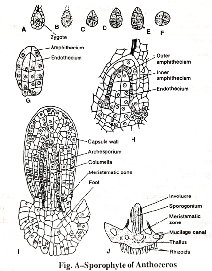
2. The Meristematic zone : A region corresponding to the seta is located between the base of the capsule and the foot. This intermediate zone is meristematic and continuously adds new cells to the tissue of the capsule. The derivatives formed by this zone progressively differentiate into columella, sporogenous tissue and wall of the capsule and finally merge with the tissue at the capsule base. It continuously grows and increases the height of the capsule even after the liberation of spores and elaters. The growth, however, is stopped after the foot is completely finished. Thus the capsule is always in different stages of growth; it is in embryonic stage at the base and mature at the tip.
3. The Capsule : The capsule forms a major part of the sporophyte. It is a slender, smooth, upright structure nearly of uniform thickness except at the slightly tapering apex. It consists of the following structures : (i) Capsule wall: It consists of 4-6 layers of parenchymatous cells. The cells of the outermost layer (epidermis) are vertically elongated with their outer walls strongly cutinised. The epidermis is interrupted at places by linear stomata each of which bears a small pore surrounded two elongated guard cells. The stomata opens in the intercellular spaces of the cells of the inner region of the wall each of which is provided with chloroplasts. The capsule wall being photosynthetic makes the sporophyte partly independent except the water and mineral nutrients for which it still depends on the tissue surrounding the foot.
(ii) Sporogenous tissue : Just beneath the capsule wall is a cylinder of sporogenous tissue series from single layered archesporium at the base, to the spores and pseudo-elaters at the tip. A progressive transformation of the sporogenous tissue into the spores and pseudo-elaters through spore mother cells and elater mother cells can be seen in the same capsule from the base upwards.
(iii) Columella: The centre of the capsule is taken up a solid column of sterile tissue called columella. It starts from the base and extends nearly to the lid. The cells of columella are narrow, elongated with more or less uniformly thickened walls. Arranged in 16 vertical rows they give the columella a shape of solid square in transverse section. The columella provides the mechanical support to the delicate capsule, helps the spores to disperse and is also said to be associated with the conduction of water.
The developing sporophyte is protected by a sheath known as involucre. It grows upwards for sometime and protect the developing sporophyte. But later on due to rapid growth of the sporophyte, the involucre ruptures and remains at the base of the sporophyte as sheath.
Evolutionary Significance of Sporophyte of Anthoceros: The sporophyte of Anthoceros exhibits a probable progressive line as follows:
1. The presence of elaborate photosynthetic mechanism (the presence of chloroplasts in capsule wall) with stomata suggest the physiological independence of sporophyte.
2. The establishment of well-developed columella suggests the beginning of formation of conducting strand.
3. The discontinued formation of sporogenous cells by the growth of sterile cells show the beginning of the formation of sporangia. zone indicates the long
4. The presence of intermediate meristematic and continued growth of sporophyte.
Anthoceros sporophyte is thus highly advanced due to the physiological independence of the sporophyte. It resembles to the sporophytes of some primitive pteridophytes e.g. Psilophytales.
Follow on Facebook page – Click Here
Google News join in – Click Here
Read More Asia News – Click Here
Read More Sports News – Click Here
Read More Crypto News – Click Here