Write an illustrated account of the progressive sterilization of sporogenous tissue among the bryophytes studied by you.
Q. Write an illustrated account of the progressive sterilization of sporogenous tissue among the bryophytes studied by you.
Ans. The production of sterile cells in the sporogonium, i.e. sterilisation of sporogenous tissue in bryophytes is quite fascinating. It is seen as a progressive conversion of simple forms of Marchantiales to complex forms of Bryales. Bower believed that the complex sporogonium of Funaria (Bryales) has evolved from the simple sporogonium of Riccia by means of progressive and ever increasing sterilisation of potentially sporogenous cells.
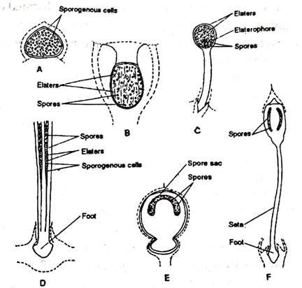
The simple type of sporogonium is seen in Riccia (Fig. A). It divides periclinal at an early stage of 20-30 celled embryo and produces outer amphithecium and inner endothecium. The whole endothecium develops into sporogenous cells while amphithecium forms protective jacket layer. Thus most of the part of sporogonium produces spores and the amount of sterile cells (jacket) is almost negligible. Occasionally some cells become converted into nurse cells and take the nutritive function. However, elaters are absent and the nurse cells are considered as the forerunners of elaters.
In Sphaerocarpus, the nurse cells are well established and in Targionia half of the sporogenous cells get converted into spores and rest half into elaters, the sterile cells. The course of evolution passing through them reaches Marchantia.

Fig. Various types of sporogonia found in the members of bryophytes (A- Riccia, B-Marchantia, C-Pellia, D- Anthoceros, E- Sphagnum, and F- Funaria
Marchantia is much advanced in that the zygote divides transversely to form a lower hypobasal cell and an upper epibasal cell. The foot and seta (sterile part of sporogonium) develop from hypobasal cell whereas capsule develops from epibasal cell. Thus half of the embryo produces sterile cells directly and does not contribute to spore producing cells. The rest half contributes to capsule. Sooner, the capsular portion differentiates into amphithecium and endothecium. The amphithecium produces one cell thick protective sterile jacket and the endothecium, produces spores as well as elaters. The elaters are single-celled and possess spiral thickenings. Thus foot, seta, jacket layer and large number of elaters are the sterile cells.
Pellia represents a still more elaborated form showing further progress in the sterilisation of the sporogenous cells. The zygote divides transversely into two cells of which lower cell contributes to foot and seta and upper cell to capsule. Thus, the 3/4 part is completely sterilised. In capsule, the sterile cells are jacket cells, elaters and elaterophore. The elaterophore, a massive sterile tissue, is found attached at the base of capsule. Thus, sterile cells are basal cell and cells of foot, seta, capsule wall, elaters and elaterophore. The presence of elaterophore leaves a clue to the beginning of columella.
In Frullania, besides foot, seta and sterile jacket cells, almost half of the cells of capsule develop into elaters and elaterophore and half into spores.
Among the members of Anthocerotales, the sterilisation proceeds ahead to that of Pellia and Frullania, where only 1/3 portion differentiates into capsule and only a smaller portion of capsule is fertile. In capsule, the amphithecium divides periclinally forming outer jacket of capsule and inner archesporium which is entirely different from that of Hepaticopsida. It is derived from amphithecium. The whole endothecium develops into central sterile cells called columella. In Anthoceros, the sterile tissues are bulbous foot, several layers of capsule wall, central columella and pseudoelaters. The outermost layer shows marked advancement. The jacket is more than one cell thick and differentiates into outer most layer epidermis, possessing chlorophyll in their cells. Stomata are also present in epidermis (fig. D).
In Sphagnum, first, there is found 5-12 celled filamentous embryo of which few upper cells develop into capsule, while middle cells form foot and seta. The lower portion of filament forms haustorium. In capsule, the entire endothecium develops into columella whereas amphithecium divides to produce outer sterile jacket and inner archesporium (sporogenous cells). The sporogenous cells arch over the columella and are arranged in 2-4 layers, i.e. only a very little portion forms spores (Fig.)
In Funaria, the sterilisation reaches to its peak where the major portion of capsule is much more complex and sterilised. The seta is well developed with ill defined internal differentiation in cell sizes. The capsule consists of several well developed sterile regions such as operculum, peristome, apophysis, columella, capsule wall of chlorophyllous cells and epidermis with stomata.
The increased amount of sterilisation increases the capacity of adaptation to the terrestrial habitat of the plant. In Bryophytes, Riccia with insignificant and negligible sterile cells is the beginning point of sterilisation which culminates into Funaria where fertile cells are insignificant in amount. Thus, Riccia and Funaria are two extremes.
Follow on Facebook page – Click Here
Google News join in – Click Here
Read More Asia News – Click Here
Read More Sports News – Click Here
Read More Crypto News – Click Here