Describe briefly the systematic position and salient features morphology and anatomy (reproductive part) of Rhynia.
Q. Describe briefly the systematic position and salient features morphology and anatomy (reproductive part) of Rhynia.
Ans. Systematic Position: Pteridophyta
Division – Psilophyta
Class – Psilophytopsida
Order – Psilophytales
Family – Rhyniaceae
Genus – Rhynia
Salient features of Morphology:
Till now only two species of Rhynia named as Rhynia major and R. gwynne-vaughani have been described by Kidston and Lang (1917, 1921) from the Old Red Sandstone Beds of Middle Devonian. As the genus has been derived from the place Rhynie situated in Scotland and, therefore, it is named after the name of the place of its origin and known as Rhynia. A number of specimens of these two species have been found at Rhynie. The specimens are so well preserved that they give detailed information about the form and structure of this very primitive vascular plant.
Kidston and Lang, thought and supported by evidences that in those times the plants grew in swampy marshes near the volcanoes.
Both the species of Rhynia, i.e., R. major and R. gwynnevaughani were herbaceous plants. Comparatively the Rhynia major was larger in size. The plant body of both the species consisted of dichotomously branched horizontal rhizome and the erect aerial dichotomously branched stem. The aerial stems of R. major were about 50 cms. high and 1.5mm. to 6mm. in diameter, whereas, the corresponding structures of R. gwynne-vaughani were 20cms. in height and 1 to 3 mm. in diameter. The aerial branches were leafless shoots. The plants were rootless. Instead of the roots, the unicellular rhizoids were present in patches on the underside of the rhizome. The aerial branches were naked, leafless, cylindrical, dichotomously forked and tapering at their apices.
The terminal, elongated sporangia were found on the tapering vegetative apices.
The stems of R. gwynne-vaughani bore adventitious branches at the bases of aerial shoots and rhizome. It is thought that these branches got detached readily from the parent plant and helped in vegetative propagation.
Anatomy:
The internal structure of the rhizome and aerial stem was quite similar to each other. This was differentiated into epidermis, cortex and stele. The details of the structure were as follows:
1. Epidermis: The epidermis was one cell in thickness with well-defined thick outer wall and conspicuous cuticle on the outer side. The cells of the epidermis were broadly fusiform from the surface view. The aerial stems bore the stomata on the epidermis here and there with pairs of guard cells. However, the rhizome did not possess any stomata. The rhizome possessed unicellular rhizoids on its surface.
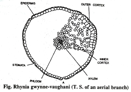
2. Cortex The cortex consists of two zones (a) the outer cortex (hypodermis) and the (b) inner cortex.
(a) Outer Cortex or Hypodermis : Immediately beneath the epidermis there was a zone one to four cells in thickness consisted of angular parenchymatous cells having no intercellular spaces known as hypodermis. This zone was also known as the outer cortex.
(b) Inner Cortex: Inner to the hypodermis there was broad zone of inner cortex. In comparison of the central stele the cortex was ten fold. The inner cortex consists of rounded cells separated from each other by large intercellular spaces which communicate to the stomata through the gaps in hypodermis. It is also believed that this zone of the inner cortex acted as the chief photosynthetic tissue.
The endodermal and pericyclic layers were altogether absent.
3. Stele: There was a protostelic type (haplostele) of stele. The central xylem core was completely surrounded by phloem layer four or five cells in thickness. The xylem consists of tracheids only. In majority of the specimens the xylem had imperfectly preserved. According to Kidston and Lang (1917), the tracheids had annular thickenings at their walls. Kidston and Lang (1920) have reported that in some of the specimens the tracheids were isodiametric, whereas in some other specimens the central tracheids were smaller in diameter and the peripheral ones larger in diameter.
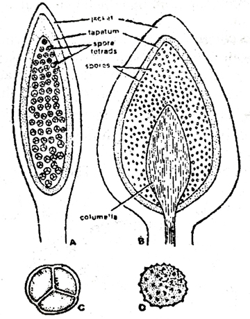
Fig. Rhynia gwynne-vaughani- A. Longitudinal section of sporangium; B. L.S. of sporangium of Hornea; C. spore tetrad of Rhynia; D. single spore of Rhynia. The phloem consists of elongated thin walled cells with oblique end walls.
4. Reproductive Structures (The sporangia): The elongated or oval sporangia were terminally developed at the apices of the branches of the aerial shoot. It is not clear whether the sporangia were borne on all the apices of the branches or on certain branches of the plant. Each apex bore a single sporangium.
As regards the shape, the sporangia were cylindrical and they possessed the greater diameter than those tips of the branches on which they were found. The jacket layer of the sporangium consists of several cells in thickness. The outermost cells of this layer were thick-walled and cutinized at their outer face. The middle layer or the sporangial wall possessed thin walled cells and inner most layer consists of thin walled rounded cells which probably act as tapetal layer. The sporangium had no columella with in it. The cavity contained many spores of the same size. The sporangia were indehiscent and it is thought that the spores were released only on the decay and disintegration of the thick sporangial wall.
The spores are spherical and cuticularized. They are found arranged in spore tetrads.
Follow on Facebook page – Click Here
Google News join in – Click Here
Read More Asia News – Click Here
Read More Sports News – Click Here
Read More Crypto News – Click Here