Describe the morphological nature of the spike of Ophioglossum.
Q. Describe the morphological nature of the spike of Ophioglossum.
Ans. Morphological Nature : According to present considerations, the fertile spike is regarded pinna-like in nature. It has been held by Goebel (1915), that the spike represents a single pinna, but according to Roeper (1859), the spike represents two basal pinnae which have fused laterally, during the ontogeny to form the spike. The interpretation is supported by the course of vascular supply to the spike. The spike exhibits the same vascular supply as would ordinarily go to a pair of pinnae.
According to Zimmermann (1930), the fertile spike and the sterile lamina are the divisions of a leaf, representing a dichotomy.
In Ophioglossum, the spike begins to grow as a small and conical outgrowth of meristematic tissue at the base of the young leaf of the adaxial side. Soon a pyramidal apical cell becomes prominent in this hump of meristematic tissue. This apical cell has four cutting faces. The apical cell starts cutting segments along its four sides that results in the formation of four quadrants. These quadrants are oriented in such a way that two lie in a plane perpendicular to the sterile lamina, and two in a plane parallel to it. In the later stage, a vertical strip of cells becomes differentiated in the epidermal layer of the two quadrants that lie in the plane parallel to the leaf blade. Each such strip is two to three cells broad and several cells in height and is called sporangiogenic band (Fig. A). Later, the hypodermal region of the band becomes differentiated into alternate blocks of archesporial cells and sterile cells (Fig. B). Each such group of archesporial cells will be a future sporangium. The archesporial cells undergo repeated divisions, resulting in the formation of a large number of sporogenous cells (Fig. C). The cells of the band which lie external to archesporial group divide periclinally to form the wall of the sporangium which is multilayered (Fig. D).
Bower (1908) described that an ill-defined tapetum of several layers of cells is formed from the outer cells of the sporogenous tissue. But now it is held that it comes from the innermost layer of wall cells. As the sporogenous cells mature, the tapetum breaks up and its cytoplasm forms a plasmodial fluid in which spore mother cells float (Fig. E).
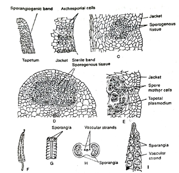
Fig. Ophioglossum vulgatum. A-E-stages of development of sporangium. F-single sporangiferous spike, G- a portion of the spike, H- transverse section of fertile spike, and I- portion of fertile spike
Most of the spore mother cells undergo reduction division to form tetrads of haploid spores (Fig. H). Each sporangium produces a very large number of spores, sometimes, this number reaches upto 10,000.
Structure of sporangiferous spike: The spike is a simple, more or less cylindrical and stalked structure (Fig. F). The fertile spike bears two marginal rows of sunken and laterally fused sporangia. The apical region of the spike which is somewhat conical and devoid of sporangia projects as a sterile process. The length of the spike varies according to the species. The number of sporangia in each spike ranges from six to twenty, according to the size of the plant.
A number of vascular strands run longitudinally up the middle and from these strands develop the lateral branches which lead to the sporangia (Fig. I.)
Follow on Facebook page – Click Here
Google News join in – Click Here
Read More Asia News – Click Here
Read More Sports News – Click Here
Read More Crypto News – Click Here